
A wrong turn near a facial nerve can turn a routine extraction into a lifelong problem. Complex tooth extractions used to carry a level of guesswork that made both dentists and patients nervous, especially when a nerve sat close to the roots. That guesswork has shrunk dramatically thanks to 3D imaging, and this piece walks through exactly how a CBCT scan dentist in Irvine relies on to plan safer, more predictable surgeries.
Seeing Beyond the X-Ray Before an Extraction
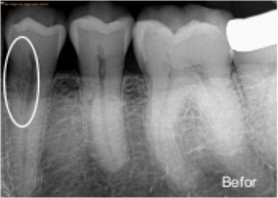
For decades, dentists planned extractions using flat, two-dimensional X-rays. Those images work fine for simple cases, but they flatten a three-dimensional jaw into a single plane. That’s a problem when a tooth root sits right next to a nerve canal, because a flat image can’t show depth or which side of the root the nerve actually sits on.
Cone beam imaging solved that blind spot. It captures the jaw in full three-dimensional detail, letting a dentist rotate the image and view the tooth from any angle. Suddenly, the exact distance between a root tip and a nerve canal becomes measurable instead of estimated.
This shift matters most with lower wisdom teeth, since their roots frequently sit close to the inferior alveolar nerve running through the jawbone. Studies on impacted third molars have shown this nerve sits right beside the tooth root in the majority of cases, and direct contact with the root happens in a smaller but still meaningful share of patients. Older imaging methods simply couldn’t tell dentists which category a specific patient fell into.
Why Nerve Proximity Is Such a High-Stakes Detail
Nerve injury during extraction isn’t common, but when it happens, the effects can linger. Numbness, tingling, or altered sensation in the lip, chin, or tongue can last months, and in rare cases, longer than that. Research tracking these outcomes has found temporary nerve symptoms after wisdom tooth removal happen in a small percentage of surgeries, while permanent symptoms are far rarer.
Knowing this risk exists ahead of time changes how a dentist approaches the entire surgery. A tooth with a nerve sitting millimeters away might call for a different extraction angle, a modified surgical technique, or even a referral to a specialist with more experience in complex cases. None of that planning is possible without clear, dimensional imaging first.
What a 3D Scan Actually Shows the Dentist
A flat X-ray gives a shadow. A 3D scan gives a map. Once a patient undergoes cone beam CT dental in Irvine imaging, the resulting scan reveals bone density, root curvature, sinus proximity, and nerve pathways all in one dataset.
That level of detail lets a dentist build a surgical plan before ever picking up an instrument. It’s the difference between walking into unfamiliar territory blind and walking in with a detailed map already memorized.
Where This Technology Shows Up Beyond Extractions
Complex extractions aren’t the only place this imaging proves valuable. Endodontic procedures like an apicoectomy in Irvine also depend heavily on knowing exactly where root tips sit relative to surrounding structures before any surgical step begins. A root that curves unexpectedly or sits closer to a sinus cavity than expected can change the entire surgical approach.
A few other situations where this imaging plays a major role include:
- Planning dental implant placement near nerve-dense areas of the jaw
- Investigating unexplained jaw pain or clicking tied to joint structure
- Locating impacted or supernumerary teeth before extraction
- Evaluating curved or unusual root anatomy before retreatment
Each of these scenarios shares one thing in common. The dentist needs depth and dimension that a flat image simply cannot provide.
How This Changes the Patient Experience
Patients often assume more imaging means more hassle, but the opposite tends to be true here. A single scan replaces the need for multiple angled X-rays and reduces the guesswork that used to require additional appointments or last-minute surgical changes. That translates into fewer surprises once the actual procedure begins.
It also means a more honest conversation before surgery starts. Rather than a dentist saying a nerve might be close, they can show a patient exactly how close it sits and explain the plan built around that measurement. That kind of clarity tends to ease a lot of pre-surgery nerves, no pun intended.
Is This Type of Imaging Right for Every Extraction?
Not every tooth needs this level of imaging. Straightforward extractions with roots nowhere near sensitive structures can often proceed safely using standard imaging alone. The decision typically comes down to what an initial exam or standard X-ray reveals about root position and nerve proximity.
Certain signs on a routine X-ray tend to push a dentist toward ordering a deeper scan rather than proceeding as usual. These include:
- Root shadows that appear to cross or overlap the nerve canal
- Unusual darkening or narrowing near a root tip
- A visibly curved or unpredictable root shape
- Interruption in the normal white outline of the nerve canal itself
Any one of these findings raises enough uncertainty that guessing simply isn’t worth the risk. A deeper scan clears up exactly what’s happening beneath the surface before any instrument touches the tooth. That extra step costs a little time upfront, but it protects both the nerve and the long-term success of the entire procedure.
Quick Answers to the Questions Patients Ask Most
Q1. Why would a dentist order a 3D scan instead of a normal X-ray?
A1. A standard X-ray flattens the jaw into two dimensions, while a 3D scan shows depth, allowing a dentist to measure the actual distance between a tooth root and a nearby nerve.
Q2. Does this imaging take longer than a regular dental X-ray?
A2. The scan itself typically takes just a few minutes, similar to other imaging visits, though results offer far more detail than a standard image.
Q3. Is the radiation exposure higher with 3D imaging?
A3. Exposure is generally low and considered safe for diagnostic use, though dentists still limit the scan area to only what’s needed for the specific case.
Q4. Can this type of scan prevent nerve injury completely?
A4. It significantly lowers the risk by revealing nerve position beforehand, though no imaging method can eliminate risk entirely during surgery.
Q5. Is this imaging only used before wisdom tooth removal?
A5. It supports many procedures beyond extractions, including implant planning, root canal retreatment, and diagnosing jaw joint issues.
Q6. How does this technology help with an apicoectomy?
A6. It shows the exact position of the root tip relative to nearby nerves and sinus structures, helping guide a safer surgical approach.
Q7. Will insurance typically cover this type of scan?
A7. Coverage depends on the specific plan and the reason for the scan, so confirming details with your provider beforehand is recommended.
Q8. How do I know if my case requires this level of imaging?
A8. Your dentist will usually recommend it after reviewing initial X-rays, particularly if there are signs the tooth root sits close to a nerve.
Clearer Images Lead to Calmer Patients and Safer Outcomes
Surgery near a nerve used to mean trusting a dentist’s estimate based on a flat picture. Today, that estimate has been replaced by an actual measurement, and that shift has quietly made complex extractions far less unpredictable than they once were.
We rely on this exact imaging approach at Irvine Endodontics because our patients deserve a plan built on real anatomy, not guesswork. While looking for a CBCT scan dentist in Irvine, you can expect the same level of detail behind every treatment plan our team builds. Hence, we’d like the opportunity to show you exactly what your scan reveals and how it shapes our approach to your care.