Root canals most often occur under a series of methods used to clean the inside of the pulp from infection and decay, and the goal of root canals is to create a tight seal, filling the canals of the tooth with an inert filling material. Like any primary root canal treatment, endodontics uses many techniques to create the tight seal needed for a successful treatment. For patients curious about their treatment, we’re here to expand on those techniques and give you a greater insight into how endodontics works within the canal system to achieve a pain-free tooth treatment.
What is Obturation in Endodontics?
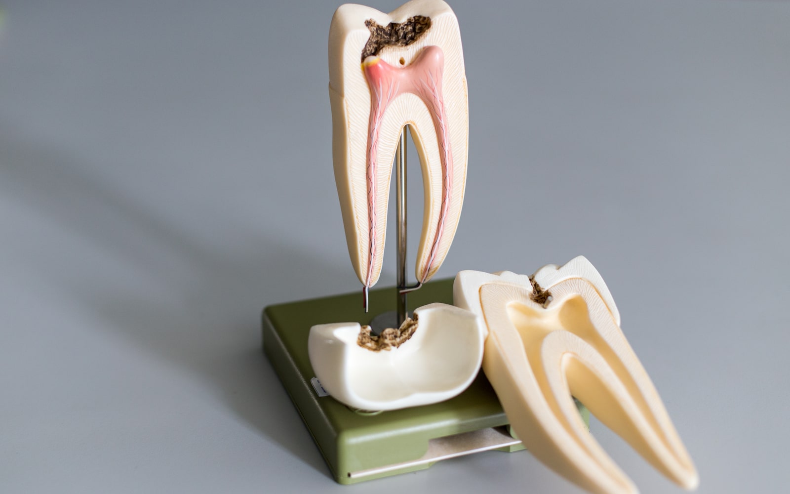
Within endodontics, many dentists are highly skilled in obturation. Obturation is described as the method used to fill and seal a cleaned root canal, using root canal filling material and sealers to complete a root canal procedure. While on the surface, the definition sounds simple, obturation is considered to be an ever-evolving art form by many in dentistry due to the complexity of the root canal system, the selective choice in materials, and most of all, the techniques used to access the root canal and apply the materials inside the tooth.
The ultimate end goal of endodontic treatment is to create successful retention to help the tooth function long term. With obturation techniques constantly advancing, clinicians must establish methods that consider the patient’s medical history and handle challenges ahead. With this in mind, the purpose of obturation is to:
- Achieve a three-dimensional tight seal within the root canal
- Prevent the development of bacteria and body fluid within the canal
- Remove bacteria and body fluid from the canal
- Prevent bacterial micro-leakage
- Replace the empty root canal space with filling material to prevent infection
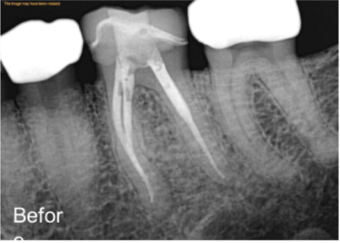
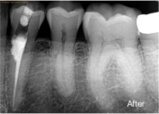
Before beginning the obturation process, endodontics must diagnose and analyze the tooth before and during treatment. In many cases, root canal treatments are appropriate for pulp exposure, apical periodontal, pulp necrosis, and irreversible pulpitis. Common tooth conditions that affect the endometrium, such as trauma, cavities, tooth loss, and improper restorations, often precede damage to the pulp and root channels.
As a general rule, endodontists will perform x-rays of the affected tooth to observe the root channels and pulp in terms of damage. Many root canal treatments vary case by case due to how the root channels develop, what access points are being affected, and where the best entry point is to provide treatment.
What Obturation Techniques Are There For Root Canals?
In many complex situations where the tooth is infected or damaged, these obturation techniques are most commonly used to provide the necessary irrigation needed for treating affected teeth:
- Cold Lateral Compaction: As the most commonly taught technique, cold lateral compaction uses scalpel blades, spreaders, and gutta-percha cones to replace defects related to the lateral or side areas of the root channels. Sealers and gutta-percha work in these cases to entomb any remaining infection that cannot be thoroughly accessed. Lateral canals make up between 27 and 45% of people’s teeth, making them difficult to irrigate during root canal therapy, making this technique a highly reliable method.
- Warm Vertical Compaction: Most often, the complex branching of root channels can sometimes lead to channels that have more vertical development, meaning they’ll firm up and down along the inside of the teeth. Warm vertical compaction attempts to compensate for damages to the vertical channels by using irrigating systems such as saline, gutta-percha cones, and heating devices that allow the gutta-percha cones to establish a tighter seal within the root channels and pulp areas.
- Warm Lateral Compaction: Like warm vertical compaction, but instead of accessing the root channels through the top of the tooth or the tooth’s grooves, it instead accesses the root channels through a lateral or side position. The same methods are applied, using sealers, irrigation acids to remove microscopic bacteria, gutta-percha cones, and gutta-percha heating devices.
- Continuous Wave Compaction: This technique is known to be less time-consuming than the warm compaction techniques. The continuous wave compaction technique uses electrical heat pluggers that provide a continuous wave of heat to the area, allowing the gutta-percha cones to have more mobility through the root channels and accelerating the packing process. Through this device, the gutta-percha is moved through the channels in a continuous motion, allowing the sealer to adhere to the inner channels of the tooth and create a tighter seal overall.
- Thermoplasticized Gutta-Percha Injection: Another modern gutta-percha delivery system, this method uses thermoplasticized gutta-percha, a modified version of gutta-percha that removes the problem of lateral condensation, or when the gutta-percha gathers and hardens along the sides of the root channels instead of fully flowing through the root channels in a complete motion.
- Carrier-based Gutta-Percha: For root channels with curvatures that exist along with the mid-root areas and coronal or front areas. This technique uses a filling technique that warms up and places the gutta-percha through a carrier device. It helps improve the fluid filtration of the channels, prevents apical leakage, and gets to the ends of the root channels, making it just as effective as lateral compaction techniques.
- ThermaFill Thermoplasticized Technique: ThermaFil, introduced in the later 1980s, allowed endodontists to access curved, long root channels with thermoplasticized gutta-percha. The Thermal brand provides their carriers with longitudinal grooves to increase flexibility and access to these areas without smearing or causing unnecessary friction when applying the gutta-percha.
- Chemically Plasticized Gutta-Percha: Instead of using carriers or heating devices, chemical solutions such as chloroform, xylol, or eucalyptol to plasticize the gutta-percha. This method is used for root channels with unusual curvatures and removes the need for heating devices to apply and seal the root channels after irrigation.
- SimpliFill Sectional Obturation: Primarily used for root channels affected along the front or coronal areas, the sectional obturation technique by SimpliFill, removing the need to make post space preparation for the root canal treatment. This technique aims to create an apical seal along the root channels that prevented leakage of the gutta-percha and lateral condensation.
- Custom Cone Obturation: Custom cone techniques work with immature root canals that have not fully formed laterally and vertically. Thus, this technique incorporated custom gutta-percha cones to accommodate for that immaturity. These custom cones can be applied to any previously mentioned techniques for a more successful root canal treatment.
- McSpadden Thermomechanical Compaction: Thermomechanical compaction works to remove the problem of temperature elevation often found with thermoplasticized gutta-percha techniques and instead uses higher speeds of instrument rotations to speed up the root canal process and prevent the problems of apical leakage of the gutta-percha. However, this method may also cause temperature rises, affecting the quality of the apical seal needed for successful root canal techniques.
Overall, endodontic techniques vary, but all are vital to the success of root canal treatments. Obturation techniques have to take in many of the clinical considerations of the patient’s tooth, their current trauma, and the extend of the damage.