A regular dental X-ray shows a flat image. A cone beam CT scan shows the full picture, every root, every nerve, every hidden problem, in three dimensions. This technology has changed how dentists and specialists plan treatment. This blog explains what a CBCT scan actually reveals, why it matters for your dental health, and when your dentist might recommend one.
Why Early Detection Depends on What You Can’t See in 2D
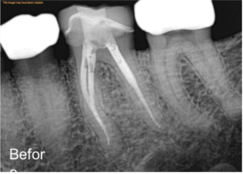
A standard dental X-ray misses up to 40% of periapical lesions, which are infections at the root tip, that a cone beam CT scan catches clearly. That’s not a small gap. It’s the difference between catching a problem early and finding it only after it’s gotten significantly worse. A flat image gives a flat picture.
A CBCT scan gives depth, detail, and a complete view of everything happening beneath the surface of your gums. For anyone exploring cone beam CT dental in Irvine, knowing what this scan actually reveals is the first step toward understanding why it’s quickly becoming a standard part of advanced dental care.
Why Flat X-Rays Sometimes Tell Only Half the Story
Traditional dental X-rays have been used for over a century, and they still have real value. They show cavities, bone levels, and general tooth positioning quickly and affordably. The limitation is that they compress a three-dimensional structure into a two-dimensional image. Think of it like trying to understand a building by looking only at its shadow. Some details show up clearly. Others disappear entirely.
A curved root tip hiding behind another root, a hairline fracture running vertically through a tooth, or a tiny infection forming along a side canal, these are exactly the kinds of things a flat X-ray struggles to show. Next comes the real problem: treatment decisions made from incomplete images can lead to incomplete results.
What a Cone Beam CT Scan Actually Does
A cone beam CT scan works by rotating a cone-shaped X-ray beam around the patient’s head. During that rotation, it captures hundreds of images from different angles. Software then combines all those images into a single, detailed three-dimensional model of the teeth, roots, jaw, and surrounding bone.
The entire scan takes less than a minute. There’s no discomfort, no needles, and no need to hold awkward positions for long. The patient simply sits or stands still while the machine moves around them. The result is a highly detailed picture that shows structures from every angle, top, bottom, front, side, and everything in between.
The Hidden Details a CBCT Scan Brings to the Surface
This is where the technology truly separates itself from traditional imaging. A cone beam CT scan reveals things that would otherwise stay hidden until they become serious problems.
For example, it shows the exact number and shape of root canals inside a tooth, which varies more than most people realize. Some teeth have two canals where a dentist might expect one. Some have unusual curves that could complicate treatment if not spotted in advance.
In addition, CBCT imaging shows the precise location of infections, the thickness of surrounding bone, and how close a root tip sits to a nerve or sinus cavity. For a procedure like apicoectomy in Irvine, this level of detail isn’t just helpful. It’s what makes the surgery safer and more predictable.
How CBCT Imaging Changes Treatment Planning
When a specialist can see the full picture before touching a tooth, the entire treatment plan improves. There are fewer surprises during procedures. The specialist knows exactly how many canals to treat, how deep to go, and how to avoid nearby structures like nerves and sinuses.
For complex cases, such as retreating a previously failed root canal, planning a surgical procedure, or evaluating an implant site, CBCT imaging is often the deciding factor in getting the outcome right. It also helps patients understand their own situation better.
Looking at a 3D image of your own tooth and seeing where the infection sits or where the fracture runs makes the treatment recommendation easier to understand and trust.
When a Dentist or Endodontist Recommends a CBCT Scan
Not every dental visit needs a cone beam CT scan. A routine cleaning or simple filling doesn’t require one. But certain situations make CBCT imaging genuinely valuable:
- Persistent tooth pain that doesn’t have an obvious cause on a standard X-ray
- Suspected root fractures that flat imaging can’t confirm
- Failed root canal treatment, where the exact cause of failure needs to be identified
- Pre-surgical planning for apicoectomy or implant placement
- Unusual root anatomy was suspected before starting root canal treatment
- Infections near sinuses or nerves, where the margin for error is small
In each of these cases, the scan gives the specialist the clarity needed to act confidently rather than making educated guesses from limited information.
What the Scan Reveals About Your Jawbone
Most people focus on what CBCT scanning shows about teeth, but the jawbone information it provides is equally valuable. The scan measures bone density, shows where bone has been lost due to infection or gum disease, and maps the exact position of the inferior alveolar nerve, which runs through the lower jaw. This nerve mapping is critical for lower molar procedures and implant placement.
In addition, CBCT imaging can reveal cysts, tumors, or other abnormalities in the jaw that would never appear on a standard X-ray. Catching these findings early, before symptoms develop, is exactly the kind of advantage this technology offers.
Radiation and Safety: What Patients Often Wonder About
A common concern patients bring up is radiation exposure. It’s a fair question. A cone beam CT scan does use more radiation than a standard dental X-ray, but significantly less than a medical CT scan used in hospitals. Most dental CBCT machines are designed to limit the beam to only the area being imaged, which reduces exposure further.
For a CBCT scan, a dentist in Irvine, following safety protocols means using the lowest effective dose for each specific case. The diagnostic benefit of catching a hidden infection, a fracture, or a nerve-adjacent root tip almost always outweighs the minimal radiation involved.
Real Questions, Straight Answers: Everything You Want to Know About CBCT Scans
Q1. Is a cone beam CT scan the same as a regular CT scan?
A1. No. A cone beam CT scan is designed specifically for dental and facial structures. It uses a focused, cone-shaped beam that rotates around the head, producing detailed 3D images of teeth, roots, and jaw. A medical CT scan covers a much larger area and uses significantly more radiation. CBCT is optimized for dental use.
Q2. How long does a CBCT scan take?
A2. The actual scanning process takes less than a minute. Including setup and positioning, most patients are done in under five minutes. There’s no recovery time needed, and you can return to normal activities immediately after.
Q3. Does a CBCT scan hurt?
A3. Not at all. The scan is completely non-invasive. You simply sit or stand still while the machine rotates around your head. There’s no contact with the teeth or gums during the scan itself.
Q4. Will my dental insurance cover a CBCT scan?
A4. Coverage varies depending on your plan and the reason for the scan. Many insurance providers cover CBCT imaging when it’s medically necessary for a specific procedure. It’s best to confirm with your provider before the appointment.
Q5. Can a CBCT scan detect all types of tooth fractures?
A5. CBCT imaging is significantly better at detecting vertical root fractures and complex cracks than standard X-rays. However, very fine surface cracks may still be difficult to see on any imaging. Your endodontist will combine scan findings with clinical examination for a complete picture.
Q6. How is CBCT imaging used for apicoectomy planning?
A6. Before an apicoectomy, the endodontist needs to know the exact position of the root tip, the size and location of any infection, and how close the area sits to nerves or sinuses. CBCT imaging provides all of this information in three dimensions, making the surgical plan far more accurate than what a flat X-ray allows.
Q7. Can children get a CBCT scan?
A7. Yes, CBCT scans can be used for children when clinically necessary, particularly for evaluating developing teeth, jaw growth, or impacted teeth. The dose is carefully adjusted, and the scan is only recommended when the diagnostic benefit clearly justifies it.
Q8. Is CBCT imaging available at most dental offices?
A8. Not all general dental offices have CBCT equipment, as it requires significant investment. Endodontic specialists and oral surgeons are more likely to have it in-house. If your general dentist recommends a CBCT scan, they may refer you to a specialist who has the technology available.
See the Full Picture Before Any Treatment Begins
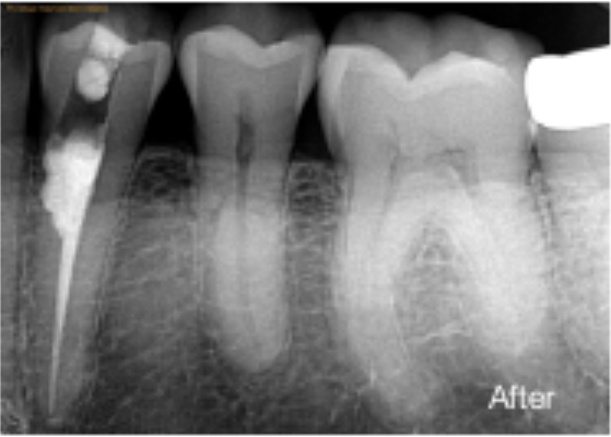
Dental treatment based on incomplete information leads to incomplete results. A cone beam CT scan removes the guesswork and gives both the patient and the specialist a clear, three-dimensional view of what’s actually happening beneath the surface. It catches what flat images miss, guides procedures with real accuracy, and ultimately helps protect the long-term health of your teeth and jaw.
Irvine Endodontics uses advanced CBCT imaging as part of a thorough diagnostic process, ensuring every treatment decision is backed by the clearest possible image. From complex root canal cases to surgical procedures like apicoectomy in Irvine, the team at Irvine Endodontics applies this technology to plan more intelligently, treat more effectively, and deliver the best possible outcome from the very first scan.