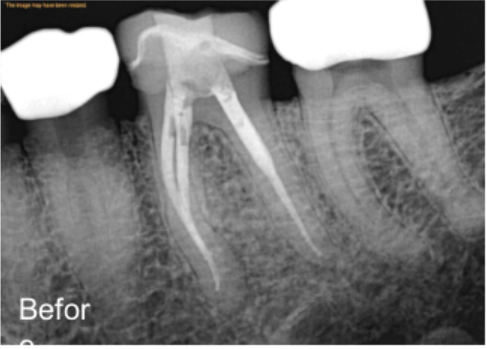
A back molar is not just a big tooth. It is a complex structure that can hold three, four, or even more canals, each one needing to be cleaned, shaped, and sealed completely. When one canal gets missed or is treated improperly, the whole procedure can fail. Understanding what makes molar root canal treatment in Orange County, CA, complex helps patients ask the right questions and choose the right specialist.
The Tooth Most Likely to Be Misread
Upper back molars are the most frequently treated teeth in endodontics, and also the most frequently treated incorrectly. That is not an opinion. Research consistently shows that missed canals in maxillary molars are one of the top reasons root canal treatment fails. The anatomy inside a molar is not always what it appears to be on a standard X-ray, and that gap between appearance and reality is where problems begin.
Most people picture a root canal as a simple, one-canal-in, one-canal-out procedure. For front teeth, that picture is mostly accurate. A molar is a different situation entirely. Upper first molars typically have three roots, but one of those roots, the mesiobuccal root, almost always hides a second canal inside it. That canal is called the MB2, and it has become one of the most talked-about challenges in the entire field of endodontics.
Why the MB2 Canal Changes Everything
The MB2 canal sits close to the primary mesiobuccal canal, separated by a thin wall of dentin. It is narrow, often angled sharply, and easy to miss without proper magnification.
Studies have found that in some populations, missing the MB2 canal was associated with apical periodontitis, which is an infection at the root tip, in more than 50% of affected molars. That is a significant number, and it tells you exactly how much this one hidden canal matters.
A molar treated without locating the MB2 may feel fine for months or even a few years. Then symptoms return, sometimes as vague pressure, sometimes as a full infection. At that point, the patient and the specialist are dealing with a more complicated situation than if the canal had been found and treated the first time.
What a Four-Canal Molar Actually Looks Like Inside
Picture the tooth as a building. The crown is the roof, the roots are the columns going into the ground, and the canals are the plumbing running through each column. An upper first molar usually has:
- A mesiobuccal root, which often contains two canals, MB1 and MB2
- A distobuccal root, which typically holds one canal
- A palatal root, which usually contains one large canal
That gives you four canals in a tooth, most people assume has three. Some molars go even further and present five, six, or, in rare documented cases, seven canals. Each one must be located, cleaned of infected tissue, shaped to allow proper filling, disinfected thoroughly, and sealed completely. Missing even one change the outcome.
The Tools That Make the Difference
This level of detail is not possible with the naked eye and a standard dental X-ray alone. Treating a complex molar well requires specific technology and the training to use it correctly.
- Dental Operating Microscope: A high-powered microscope gives the endodontist a magnified, brightly lit view of the pulp chamber floor, where canal openings are located. The MB2 canal is often just a subtle discoloration or a hairline groove, invisible without magnification. Studies show that microscope-assisted access improves the rate of locating MB2 significantly compared to unaided exploration.
- CBCT Imaging: Cone-beam computed tomography produces a three-dimensional image of the tooth before treatment begins. A standard X-ray flattens a three-dimensional structure into two dimensions, which means extra canals, unusual root curves, and accessory anatomy often go unseen. CBCT removes that guesswork entirely. The specialist can map every canal, every curve, and every variation before the first instrument enters the tooth.
- Nickel-Titanium Rotary Files: These flexible instruments follow the natural curve of a canal without forcing it straight, which protects the root structure during cleaning and shaping. Older stainless-steel files were stiffer and more likely to create problems in curved or narrow canals.
- Ultrasonic Tips: Fine ultrasonic instruments help remove calcified dentin blocking the entrance to a hidden canal. Calcification is common in older teeth or teeth that have experienced trauma, and it can make the MB2 even harder to find.
When Calcification Makes It Harder
As teeth age, the body naturally deposits dentin inside the canals in a process called sclerosis. This happens from the top of the canal downward. A heavily calcified canal can appear completely blocked on an X-ray, leading some practitioners to assume it does not need treatment or simply cannot be accessed.
However, a skilled endodontist with proper magnification and ultrasonic instruments can often navigate through the calcified section and locate the canal below it.
This is one of the key moments where the experience level of the treating specialist matters most. Removing calcified dentin to find a hidden canal requires a steady hand, excellent visualization, and a thorough understanding of where the canal is likely to be based on the tooth’s internal anatomy.
Irrigation: The Step That Cleans What Instruments Cannot Reach
Mechanical instrumentation cleans the main body of each canal. But a molar’s root canal system is not just a series of straight tubes. It has lateral branches, fins, and isthmus connections between canals, small passageways where bacteria can survive even after thorough filing. Irrigation protocols using sodium hypochlorite and EDTA, often activated with ultrasonic energy, reach areas that files simply cannot.
Skipping or rushing this step is another common reason root canals fail, particularly in molars, where the internal anatomy is most complex. Proper irrigation is not an add-on. It is a core part of what makes root canal microsurgery treatment in Orange County, CA, effective in difficult cases.
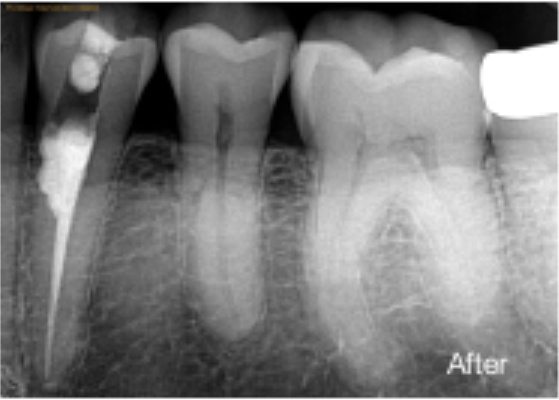
Obturation: Sealing Every Space Completely
Once all canals are cleaned and shaped, they need to be filled and sealed. Gutta-percha, a rubber-like material, is packed into each canal along with a sealer to close off any remaining space. In a molar with four canals, this step has to be done correctly in each one. A poor seal in even one canal creates a pathway for bacteria to re-enter, which can restart the infection and lead to treatment failure.
Warm vertical compaction is the most widely used technique for molar obturation because heat makes the gutta-percha flow and adapt to the irregular shapes within a complex canal system. The goal is a dense, gap-free fill from the tip of the root to the access opening, confirmed with a post-treatment X-ray before the appointment ends.
What Happens When a Canal Gets Missed
A missed canal is not a minor oversight. Organic tissue left inside an untreated canal continues to break down. Bacteria feed on it. An infection develops at the root tip, and that infection spreads into the surrounding bone. The patient may not feel it for some time, but the damage continues.
Eventually, a periapical lesion, visible as a dark shadow on an X-ray, forms around the root. At that point, the tooth often needs retreatment or, in some cases, endodontic microsurgery to resolve the infection surgically.
Research from the American Association of Endodontists confirms that endodontic microsurgery, when performed properly, achieves success rates comparable to or higher than nonsurgical retreatment in many complex situations. The key factor in both scenarios is catching the problem and addressing it completely.
Straight Answers About Complex Molar Root Canals
Q1: How do I know if my molar has four canals?
A1: You cannot know without proper imaging. A CBCT scan before treatment gives the clearest picture of how many canals your molar actually has.
Q2: Does a four-canal root canal hurt more than a regular one?
A2: No. Local anesthesia numbs the area fully regardless of how many canals are involved. The complexity affects the skill and time required, not the level of discomfort during treatment.
Q3: What is the MB2 canal and why does it get missed?
A3: It is a second canal inside the mesiobuccal root of upper molars. It is narrow, angled, and nearly invisible without magnification, which is why it gets missed frequently.
Q4: Can a missed canal be fixed after the fact?
A4: Yes. Nonsurgical retreatment or endodontic microsurgery can address a missed canal, though both are more involved than getting it right the first time.
Q5: How long does a complex molar root canal take?
A5: Most complex molar cases take 90 minutes to two hours. Some may require a second visit if the canal location or disinfection needs additional time.
Q6: Is a crown always needed after a molar root canal?
A6: Almost always. A molar takes significant biting force daily. Without a crown, the treated tooth is at real risk of fracturing, which can lead to tooth loss.
Q7: What is the success rate for complex molar root canals?
A7: When treated by an experienced endodontist using a microscope and CBCT imaging, success rates for complex molar root canals are consistently high, often above 90% in documented studies.
Q8: How is endodontic microsurgery different from a regular root canal on a molar?
A8: A standard root canal works from the crown downward. Microsurgery accesses the root tip through a small incision in the gum to remove infection that nonsurgical treatment cannot fully reach.
The Molar That Deserves to Be Treated Right the First Time
Complex molars leave very little room for error. A missed canal or incomplete cleaning can lead to reinfection years later. Successful root canal treatment in Orange County, CA, depends on treating every part of the tooth thoroughly the first time.
At Irvine Endodontics, every molar case is treated with CBCT imaging and a dental operating microscope because molar anatomy is rarely simple. We often help patients whose previous treatment missed hidden canals or calcified roots, once considered untreatable. In many cases, the tooth was still worth saving, and we saved it.